False Positive SCID Screens: Common Causes
A positive newborn screen for Severe Combined Immunodeficiency triggers a chain of events most pediatricians will navigate at least once in their career — and more often than the literature admits.
False Positive SCID Screens: Common Causes and What Drives Them
Roughly 1 in 20,000 infants will flag as having low T cells on the dried blood spot assay, but only about 1 in 58,000 actually has SCID. That gap is the subject of this article: the clinical and biological reasons a TREC-based screen returns abnormal in a child who does not have SCID, and what an experienced clinician should expect when triaging those results.
I've spent years running pediatric immunology cohorts, and I'll say plainly that the TREC assay is a remarkably specific test — its false-positive rate sits well below 1% — but "well below 1%" still translates into real newborns, real anxious parents, and real follow-up costs when multiplied across every state program. Knowing why those false positives occur is what separates a screening reflex from a diagnostic workup that wastes three weeks of a family's life.
The Mechanics of TREC-Based SCID Screening
The assay measures T-cell receptor excision circles (TRECs) — small circular DNA fragments produced during the V(D)J recombination that generates a functional T-cell receptor in naive T lymphocytes. TRECs are stable, do not replicate with cell division, and therefore serve as a proxy for recent thymic emigrants. A newborn with adequate thymic output produces TRECs in easily detectable quantities; a baby with profound T-cell deficiency produces very few or none.
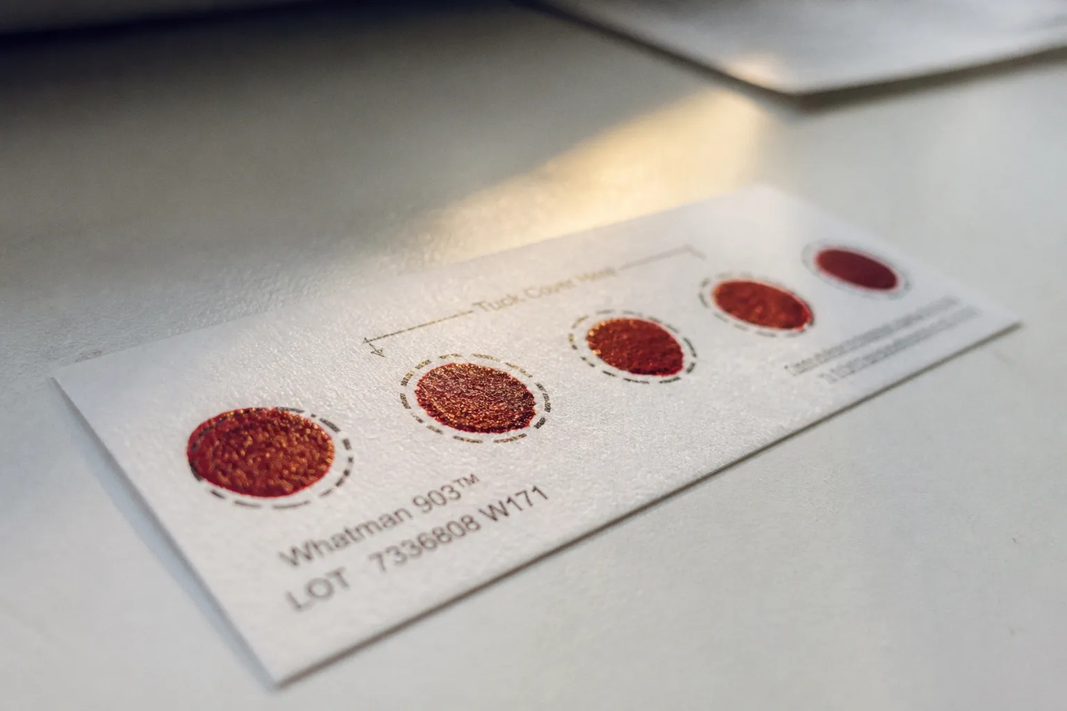
The screening pipeline is straightforward in principle. A heel-stick dried blood spot is eluted, subjected to quantitative PCR, and the TREC copy number is compared against a state-defined cutoff. If the value falls below threshold, the case is flagged and referred for confirmatory flow cytometry and, increasingly, targeted genetic sequencing. The TREC itself is not diagnostic of SCID — it is a population-level filter that says, in essence: this infant warrants a deeper look.
The critical detail is that the assay detects a signal (low thymic output), not an etiology. Anything that suppresses thymic output — even transiently — can produce an abnormal screen. That's the structural reason false positives exist, and it's the lens we should apply to every positive result that does not turn out to be SCID.
A positive TREC screen is not a diagnosis of SCID. It is a signal of low thymic output, and the differential for low thymic output in a newborn is broader than most clinicians were taught.
Impact of Gestational Age and Low Birth Weight on T-Cell Maturation
Prematurity is the single largest driver of false-positive TREC screens in any high-volume program, and it's also the one that gets mishandled most often in the early days of a state screening rollout. The underlying biology is non-negotiable: T-cell subsets and TREC values mature and increase with advancing gestational age. A 30-week infant simply has not had the thymic exposure time of a 40-week infant, and the assay picks that up.
In my experience running these cohorts, the practical consequence is that NICU graduates generate a disproportionate share of abnormal screens relative to their population share. The 2013 publication flagging diagnostic challenges in premature infants remains relevant; the 2023 multi-state evaluation reinforced the point with broader numbers. State programs have responded with gestational-age-adjusted cutoffs or repeat-screening protocols, but variability persists between jurisdictions, and a clinician receiving a positive screen on a 32-week ex-preemie should not assume the result is a clean false positive until the repeat or confirmatory flow cytometry is back.
Low birth weight compounds the issue independently. Infants under approximately 1,500–2,000 g, regardless of gestational age, tend to run lower TRECs, and the overlap with true SCID distributions is real. This is why the California cutoff for significant T-cell lymphopenia referral sits at ≤1,500 T cells/μl on confirmatory testing rather than at the TREC value alone — the confirmatory flow cytometry provides the second filter that gestational age alone cannot.
Secondary T-Cell Lymphopenia: The Role of NICU Care and Surgery
Beyond prematurity, secondary T-cell lymphopenia is the second big bucket of false positives, and it's where clinical context matters most. Several NICU-associated exposures reliably depress TREC values in newborns who do not have primary immunodeficiency:
- Neonatal intensive care admission for any reason — sepsis, respiratory distress, surgical evaluation — correlates with lower TRECs at first screen.
- Heart surgery and major neonatal surgery produce transient T-cell depletion through blood loss, transfusion-related lymphocyte dilution, and stress-mediated redistribution. The thymus is not the target; circulating T cells simply fall.
- Severe acute illness in the newborn period (viral sepsis, bacterial sepsis with lymphopenia, congenital infections) suppresses lymphocyte counts across the board.
- Prenatal corticosteroid exposure, given to mothers at risk of preterm delivery to accelerate fetal lung maturity, has been associated with altered neonatal lymphocyte trafficking and lower TREC values at the time of first screen.
The honest version of this, which I wish were more explicit in screening program communications: an abnormal TREC in a NICU graduate is often a marker of how sick the baby has been, not a marker of an intrinsic immune defect. The follow-up protocol has to allow for this. Programs that reflex immediately to genetic sequencing without an interim flow cytometry in these cases generate cost and anxiety without diagnostic yield.
| Exposure / Context | Mechanism of TREC Depression | Typical Course |
|---|---|---|
| Prematurity (<34 weeks) | Immature thymic output | Resolves with advancing age; repeat screen |
| Low birth weight (<2,000 g) | Reduced thymic mass and output | Repeat at corrected gestational age |
| NICU admission | Stress lymphopenia, illness-related | Resolves post-discharge |
| Major neonatal surgery | Transfusion dilution, redistribution | Resolves within weeks |
| Prenatal corticosteroids | Altered lymphocyte trafficking | Transient; resolves by 2–4 weeks |
| Severe neonatal infection | Sepsis-related lymphopenia | Resolves with recovery |
Syndromic Mimics: Genetic Conditions Affecting TREC Values
The third category is the one that worries me most as a clinical auditor, because these are true positives to a screening signal without being SCID. Several non-SCID primary immunodeficiencies and syndromic conditions produce abnormal TREC values and require their own diagnostic pathways:
- DiGeorge syndrome (22q11.2 deletion syndrome) — thymic hypoplasia or aplasia produces low TRECs in proportion to the degree of T-cell compromise. Partial DiGeorge with normal or near-normal T cells can produce mildly abnormal screens that normalize; complete DiGeorge presents with persistently low TRECs and requires the same urgency as SCID for immune reconstitution planning.
- Ataxia-telangiectasia — a DNA repair defect that also produces T-cell lymphopenia. The ATM gene is often included on the targeted sequencing panels deployed after a positive TREC screen, which is why this diagnosis surfaces during the workup rather than later.
- CHARGE syndrome — coloboma, heart defects, atresia choanae, growth retardation, genital anomalies, ear anomalies — is associated with variable T-cell deficits related to thymic maldevelopment.
The clinical lesson: a positive TREC screen in a newborn with congenital heart disease, characteristic dysmorphology, or a syndromic features deserves immediate genetic consultation even before flow cytometry returns. Several of these conditions have actionable management windows (live vaccine avoidance, transfusion policy, prophylactic antimicrobials) that intersect with the workup timeline.
The undetermined territory — flagged as unknowns in the research base and worth saying out loud — is the exact nationwide proportion of false positives attributable to prenatal corticosteroids versus surgery, and the precise prematurity-adjusted false-positive rate aggregated across all state programs. State-level variability is wide enough that any single number underestimates the range.
Clinical Pathways Following an Abnormal Newborn Screen
The follow-up algorithm is where the system earns or loses the screening's value. A well-run pathway looks roughly like this in my experience:
1. Immediate confirmatory flow cytometry with CD3, CD4, CD8, CD19, CD16/56, naive T-cell markers, and recent thymic emigrant quantification. This step alone resolves most premature-infant false positives and identifies syndromic T-cell lymphopenia.
2. Targeted genetic sequencing — increasingly a rapid panel covering SCID-causing genes (IL2RG, JAK3, RAG1, RAG2, ADA, DCLRE1C, LIG4, NHEJ1, AK2) plus syndromic mimics (22q11.2, ATM, CHD7). The turn-around has compressed to days in many centers.
3. Repeat TREC at corrected gestational age for premature infants with normal flow cytometry — a safety net for the small fraction whose flow is borderline.
4. Specialist referral to pediatric immunology for any confirmed T-cell lymphopenia below the program threshold, regardless of etiology.
The mistakes I see when the pathway is poorly implemented: skipping flow cytometry in favor of direct genetic sequencing (cost without benefit), failing to repeat the screen in premature infants who looked reassuring on first workup, and not communicating to families that "abnormal screen" is a triage category, not a diagnosis. Roughly 1 per 15,000 births in California crosses the threshold for significant T-cell lymphopenia referral — a non-trivial fraction of those will resolve, and parents deserve to hear that probability from a clinician rather than the lab report.
What the False-Positive Rate Actually Means
Returning to the headline number: a TREC assay false-positive rate below 1% is genuinely excellent for a population screening test. The reason it still merits a dedicated article is that, applied across approximately 3.6 million US births per year, even a quarter-percent false-positive rate produces thousands of abnormal screens annually — and the biology of those screens is heterogeneous enough that pattern recognition matters.
My practical verdict after reviewing cohort after cohort of these cases: the assay is doing exactly what it should. It is broad on purpose. The clinical system downstream of it — the flow cytometry, the genetics, the repeat screening, the immunology referral — is what determines whether the screening program's promise translates into better outcomes. Where that downstream system is well-resourced, false positives are absorbed without harm. Where it is fragmented, every false positive becomes a small clinical failure of its own.
The honest gap, and the one I would prioritize in any quality-improvement conversation, is gestational-age-adjusted protocols. They exist in some programs and not others, and they are the single most evidence-backed lever for reducing avoidable follow-up without missing true SCID. Until that variation closes, expect the prematurity-driven false positive to remain the most common category the clinician encounters — and expect the syndromic mimics to keep being the ones that justify the entire screening enterprise when caught early.