Pediatric flow cytometry: why age-specific ranges matter
California's newborn screening program pushed 3,252,156 infants through T-cell receptor excision circle (TREC) testing from dried blood spots in a single evaluation period. Every flagged sample then handed off to flow cytometry for confirmation.
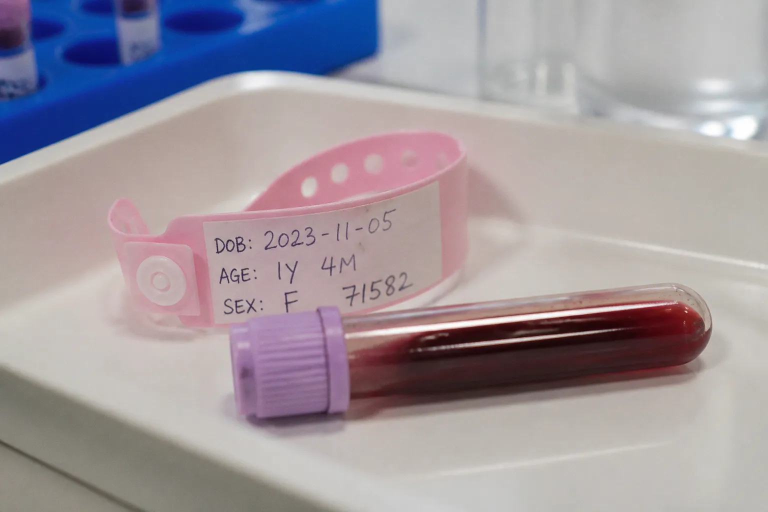
The scale tells you the operational reality: behind every SCID screen sits a pediatric immunophenotyping lab — or it should. And this is where the diagnostic machinery starts showing its seams. Adult reference ranges threaded into a neonatal cohort don't just look wrong; they misclassify at a rate the field still underestimates.
The kinetic shifts happening in a child's immune system across the first five years of life are not subtle. Total B-cell numbers, transitional B-cell counts, and the naive-to-memory lymphocyte ratio all move on trajectories that bear almost no resemblance to the steady-state distributions clinicians memorized for adult patients. Treat a 3-year-old's flow cytometry panel as if it were a 40-year-old's, and you invite false alarms, missed diagnoses, and a steady stream of referrals that don't need to exist.
The Kinetic Shift: Why Pediatric Immune Development Defies Adult Norms
The first misconception I see in requisitions and result interpretations is treating peripheral blood lymphocyte subsets as if they were baseline-stable physiological constants. They are not. The pediatric immune system is not a scaled-down adult immune system; it is a different organ undergoing active construction, with lymphocyte compartments moving on trajectories that have no analog in adult reference data.
Adult reference ranges describe a steady-state system. Pediatric reference ranges describe a system in motion.
Total B-cell numbers and transitional B-cell counts decline most rapidly between roughly 1 and 5 years of age, while memory B cells accumulate across the same window. The CD4-to-CD8 ratio sits in a different part of the distribution during early childhood than it does in adults. Even the naive-to-memory switch within the T-cell compartment is mid-transition at the exact moment most clinical laboratories are running the panel. Diagnostic flow cytometry as a modality depends entirely on the reference interval you apply to the patient sitting in front of the cytometer. Pull a value outside the adult range, and the report flags that often arrive marked "low" or "high" are stripped of any pediatric context. The instrument doesn't know the patient's age. The reference database does.
Garcia-Prat and colleagues published extended immunophenotyping reference values in 2018, and a Finnish cohort followed in 2025 establishing extended pediatric immunophenotyping reference values — both of which exist in the literature precisely because the gap between adult normals and pediatric reality was too wide to ignore. The 2019 California newborn reference interval study, published in the Journal of Allergy and Clinical Immunology through NewSTEPs, mapped effective gestational ages from 22 to 52 weeks, effectively covering the entire perinatal window. The fact that research groups are still publishing fresh pediatric reference intervals in 2025 and 2026 tells you the field is not done.
Beyond the Adult Baseline: Mapping Lymphocyte Subset Evolution
The standard adult reference ranges are not exotic. Laboratory medicine has spent decades normalizing them. The problem is that they are, by definition, not pediatric.
| Subset | Adult reference range | Pediatric trajectory (qualitative) |
|---|---|---|
| CD19+ B cells | 6–22% | Elevated in young children; declines toward adult range by ~5 years |
| CD3+ T cells | 65–85% | Lower in infancy; rises with age |
| CD4+ T helper/inducer | 30–62% | Higher in infancy; gradually normalizes |
| CD8+ T cytotoxic/suppressor | 13–37% | Lower in infancy; rises with age |
| NK cells (CD16+/CD56+/CD3−) | 5–31% | Variable; often higher in early life |
| CD4/CD8 ratio | 0.7–2.7 | Elevated in infancy; narrows with age |
The columns are not interchangeable. A 6-month-old presenting with a CD4 count that would be flagged as "low" against an adult range might be entirely within the expected distribution for that gestational-corrected age. A 4-year-old whose CD19 percentage sits above the adult ceiling may not be lymphoproliferative; they may simply be on the standard pediatric arc. Direction and magnitude of these shifts vary across published cohorts, but the directional pattern is consistent.
The clinical pitfall is structural. Whole blood lysis flow cytometry instrumentation has not changed dramatically over the past two decades — the antibodies, the lasers, the gating strategies are roughly the same. What has changed is the labor required to maintain a pediatric-appropriate reference database. Laboratories that have not invested in this work are the ones filing pediatric reports under adult intervals. The result is not subtle and not rare.
Sample handling compounds the problem. Whole blood lymphocyte flow cytometry has a stability window of roughly 24 hours at room temperature. For adult patients, this is usually workable. For pediatric patients, especially neonates in a referral network where the sample is drawn in a regional hospital and shipped to a tertiary immunology lab, the timing pressure is real. A delayed sample doesn't deliver a slightly degraded pediatric result; it delivers a delayed adult-range interpretation of a pediatric sample, which is worse than nothing.
From TREC Screening to Flow Cytometry: Bridging the Diagnostic Gap
The California cohort is the most useful single data point I have for understanding how newborn screening for SCID actually works at population scale. TREC from dried blood spots served as the first-pass filter. Three million, two hundred fifty-two thousand, one hundred fifty-six infants went through it. The infants who flagged positive then advanced to follow-up flow cytometry for lymphocyte subset confirmation.
This is the moment where the diagnostic apparatus either functions or fails. TREC screening is, in my view, a clean design — high sensitivity, acceptable false-positive rate, no need for age-specific reference intervals because the assay is a copy-number measurement rather than a relative percentage. Flow cytometry is the downstream step that introduces the age-dependence problem.
The California evaluation found that sex and ethnicity were not significant determinants of neonatal T-cell and B-cell subset counts in their cohort. That finding matters because it pulls the field's attention away from demographic adjustments and onto age stratification. The cohort ages in that study ran from 22 to 52 weeks effective gestational age — meaning the entire perinatal window was effectively sampled, and what moved the reference intervals was not the patient's demographics but their developmental stage.
Yet across most clinical laboratories operating today, the pediatric flow cytometry report still ships with adult reference ranges printed on the bottom of the page. That's a regulatory gap as much as a clinical one. The assay is validated; the platform is approved; the reagent is licensed. But the interpretive framework the laboratory attaches to the result is not pediatric calibration in most cases. It is adult calibration grafted onto a pediatric patient.
Interpreting Functional Assays: The Naive Lymphocyte Conundrum
Reference intervals are only half the problem. Functional assays — the ones that tell you whether the lymphocytes you counted can actually do their job — carry their own pediatric blind spot.
The FASCIA method for lymphocyte mitogen response testing consistently yields lower stimulation results in children than in adults. The default interpretation under adult reference expectations is that pediatric responses are suppressed. The biological reality is that children have a higher proportion of naive lymphocytes than adults, and naive lymphocytes respond less vigorously to mitogen stimulation than memory cells do. Suppressed is not the right word. Developmentally appropriate is.
A naive lymphocyte is not a defective lymphocyte. Treating the two as interchangeable is how misdiagnoses are born.
The clinical risk here is twofold. The first is the false-positive immunodeficiency workup, which generates anxiety, repeat testing, and unnecessary referrals. The second is more dangerous: a child with a genuine functional immunodeficiency whose naive-predominant blood produces a stimulation result that gets dismissed as "the normal pediatric pattern." Both errors come from the same root cause — using adult-derived functional reference data to interpret a pediatric sample.
Mitogen response panels — phytohemagglutinin, concanavalin A, pokeweed mitogen — were calibrated on adult cohorts. The pediatric dose-response curves have been established in research settings but rarely translate into clinical laboratory reference intervals. The clinician ordering the test often receives a numerical result, a reference range, and a flag. The flag is rarely age-adjusted. This is where I expect the field to move next. The California TREC experience taught the screening community that age-stratification of the downstream assay matters as much as the upstream filter. Functional assays are the next layer of the same problem.
Standardizing Pediatric Panels: Navigating Variable Reference Intervals
The standard antibody panel for evaluating pediatric and adult lymphocyte subsets is not the source of the difficulty. The same markers do the work in both populations: CD3, CD4, CD8, CD16, CD56, CD19, and CD45 make up the core of any reasonable clinical immunophenotyping order. The platform variation between instruments is real — different vendors, different fluorochromes, different compensation matrices — but the underlying biology the panel interrogates is the same.
The variability that matters is the reference interval the laboratory attaches to the panel. There is no universal global consensus on unified pediatric flow cytometry reference ranges that spans all laboratory equipment platforms and ethnicities. That is not a gap I'm projecting; it is an open question in the field, and it shows up in the fact that research groups continue to publish platform- and cohort-specific pediatric reference intervals as recently as 2025.
The practical implication for clinicians ordering the test and parents receiving the result is that the same numerical value can carry different clinical meaning depending on which laboratory produced the report. A CD19 percentage of 18% at a lab using adult reference ranges sits within range. The same 18% at a lab using an appropriately age-stratified pediatric range may sit at the upper edge of normal for a 2-year-old, or low for a 10-year-old. Observed value identical. Clinical interpretation opposite.
For laboratories that have not built or licensed a pediatric reference interval database, the operational work has several non-negotiable components:
- Build age-stratified pediatric reference intervals on the lab's own platform — borrowed validations from another vendor's matrix are not directly transferable.
- Cross-reference the new intervals against at least one published cohort (Garcia-Prat 2018, the 2025 Finnish study, or the California NewSTEPs data) before clinical deployment.
- Update the report template to display age-appropriate ranges at the result line, not buried in a generic disclaimer.
- Audit a retrospective sample of recent pediatric reports to quantify the current misclassification rate — most labs that run this exercise find a non-trivial number.
- Establish sample-handling protocols that account for the 24-hour stability window, especially for shipments of neonatal samples between regional draw sites and tertiary immunology labs.
The California NewSTEPs data, the 2019 journal publication, and the more recent cohort work all converge on a single operational message: pediatric flow cytometry requires age-specific reference intervals, and the instruments will not produce them by default. The clinical laboratory has to build them, validate them, and maintain them. Until that work is done at scale, the report carries an interpretive debt the clinician inherits.
The Sober Verdict
Pediatric flow cytometry is not a niche corner of laboratory medicine. It is the diagnostic backbone for the entire inborn errors of immunity and pediatric immunodeficiency spectrum, and it is the downstream confirmatory step for every TREC-positive newborn screen. The assay infrastructure works. The reference interval infrastructure, in most laboratories, does not.
I am not persuaded by arguments that adult reference ranges are "close enough" for clinical use in children. The data on lymphocyte subset kinetics from infancy through age 5 is unambiguous — the system is in motion, and the adult range describes a different system at a different stage. Laboratories that continue to apply adult intervals to pediatric samples are accepting a misclassification rate that has no clinical justification.
The path forward is operational, not theoretical. Clinical laboratories need to invest in age-stratified pediatric reference intervals, validate them against their specific platforms, and update them on the cadence the immune system requires — not the cadence laboratory software updates ship on. The regulatory and reimbursement frameworks will eventually catch up. The clinical question — does this child have a real immunodeficiency, or is the lab reading a moving target with a static ruler — cannot wait.