Dihydrorhodamine assay: the path to CGD diagnosis
When a child has recurrent pneumonia, deep bacterial or fungal infections, unusually persistent lymphadenitis, or granulomatous inflammation, the central clinical question is not simply whether the child is getting sick too often.
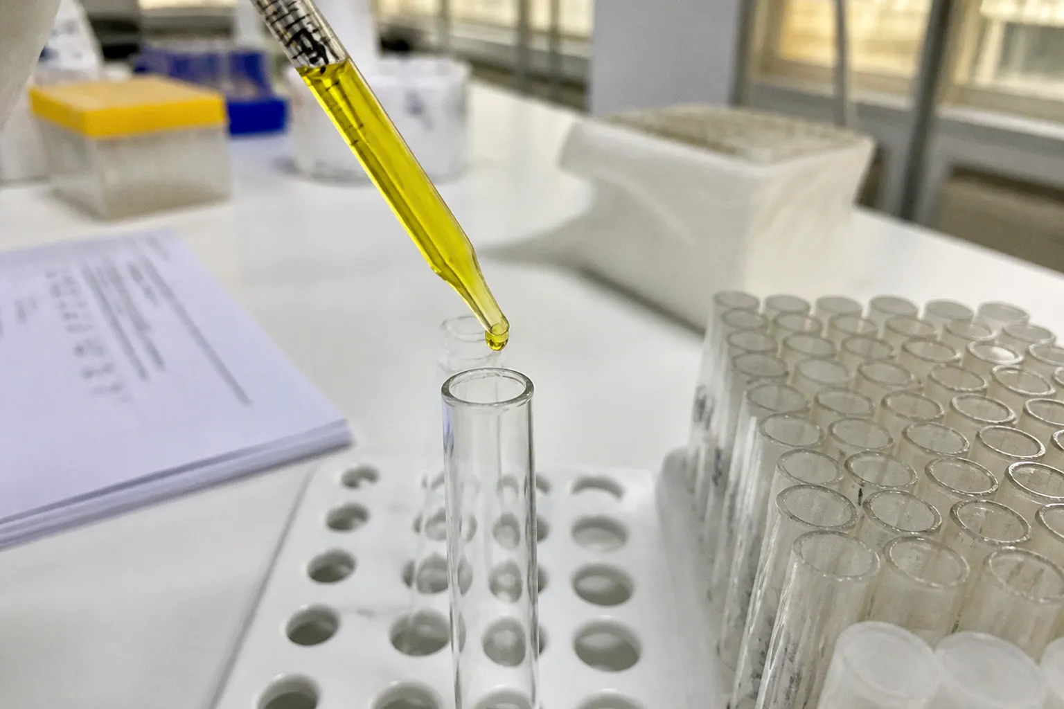
We need to know whether neutrophils can perform one of their most important tasks: generating the oxidative burst required to kill certain microbes after phagocytosis.
The dihydrorhodamine assay, usually called the DHR or DHR flow cytometry test, is the primary functional screening test for chronic granulomatous disease, or CGD. It measures neutrophil respiratory burst activity in a blood sample and can often show not only that the pathway is impaired, but also whether the pattern is consistent with an X-linked form, an autosomal recessive form, or carrier status in a female relative. For families and clinicians, this makes DHR testing a central point on the CGD diagnostic pathway in children.
A DHR result is not the whole diagnosis. It is a functional map of the immune defect, which then needs to be connected with the child’s clinical presentation, laboratory findings, and molecular testing.
Why CGD enters the differential diagnosis
CGD is an inherited disorder of the phagocyte NADPH oxidase complex. In a healthy neutrophil, this complex helps produce reactive oxygen species during the respiratory burst. These substances contribute to the destruction of bacteria and fungi that have been engulfed by the cell.
When the oxidative burst is absent or substantially reduced, neutrophils may still move toward infection and ingest microorganisms, but their intracellular killing capacity is impaired. The result can be a pattern of infections that is more invasive, prolonged, recurrent, or difficult to eradicate than expected.
The estimated global incidence of CGD is approximately one in 200,000 live births, although the observed frequency varies between populations and may be higher in communities with increased consanguinity. The condition can affect children of any sex. X-linked CGD is more common in boys because the relevant gene, CYBB, is located on the X chromosome, while autosomal recessive forms can affect both boys and girls.
The clinical presentation may include:
- recurrent bacterial or fungal pneumonia, particularly when infections are severe or respond incompletely to standard treatment;
- suppurative lymphadenitis, liver abscesses, or other deep-seated infections;
- osteomyelitis or persistent skin and soft-tissue infections;
- infections caused by organisms that are not typically invasive in otherwise healthy children;
- granulomatous inflammation involving the gastrointestinal or genitourinary tract;
- poor growth, prolonged fever, or repeated hospital admissions without a clear explanation;
- a family history of severe childhood infections or early deaths from infection.
None of these findings is specific to CGD. Many children with recurrent infections do not have an inborn error of immunity. The diagnostic value lies in the pattern, the severity, the organisms involved, the response to therapy, and the presence of inflammatory complications.
The DHR assay does not ask whether a child has had too many infections; it asks whether the child’s neutrophils can complete a critical antimicrobial function.
How the DHR assay measures neutrophil function
The test uses flow cytometry to examine individual blood cells and quantify their fluorescence after stimulation. The laboratory begins with dihydrorhodamine 123, a cell-permeant compound that is initially non-fluorescent. During the oxidative burst, hydrogen peroxide and related reactive oxygen species are generated. Dihydrorhodamine 123 is oxidized into rhodamine 123, which emits bright fluorescence.
In practical terms, the laboratory compares neutrophil fluorescence before and after stimulation, commonly using phorbol 12-myristate 13-acetate, or PMA. The PMA stimulation is performed under controlled laboratory conditions, with the supplied protocol including incubation for approximately 15 minutes at 37°C. Flow cytometry then measures the fluorescence shift across a large number of cells.
This is why DHR is considered a neutrophil function test by flow cytometry rather than a conventional antibody or cell-counting assay. The question is functional: after stimulation, do the neutrophils generate the expected oxidative response, and what does the cell population look like?
The DHR readout may reveal several clinically meaningful patterns:
- Absent fluorescence shift: this is compatible with a severe defect in the oxidative burst, such as a classic X-linked CGD pattern involving the gp91phox component.
- Markedly reduced or partial fluorescence: this may occur with residual oxidase activity or certain autosomal recessive defects.
- Bimodal fluorescence pattern: two neutrophil populations, one responding and one not responding, can suggest a carrier state for X-linked CGD in a female.
- Near-normal or mildly reduced fluorescence: this does not automatically exclude every form of CGD, particularly rare defects in which DHR performance may be less clearly abnormal.
The laboratory may report mean fluorescence intensity, stimulation indices, histograms, or other flow cytometric measures. Exact numerical cutoffs are not universal: they depend on the instrument, reagents, laboratory protocol, control sample, and reporting system. A DHR result should therefore be interpreted using the reference framework of the performing laboratory rather than by applying a cutoff found in an unrelated report.
The pediatric diagnostic steps, from suspicion to confirmation
The most reliable approach is a staged management pathway. DHR testing is usually ordered when the clinical presentation justifies a functional assessment of phagocyte activity, but the pathway begins before the blood draw.
1. Define the clinical signal
The first step is to characterize the infection and inflammation history rather than count episodes in isolation. We look at the age at onset, anatomical sites, organisms, duration of illness, need for intravenous antimicrobials, complications, and whether the child returns to baseline between infections.
The same principle applies to inflammatory disease. Granulomatous colitis, unexplained hepatic lesions, recurrent perianal disease, or persistent lymph node inflammation may be relevant even when infections have not yet been formally identified.
A careful pedigree is also valuable. X-linked CGD may present through affected boys on the maternal side of a family, while autosomal recessive inheritance can appear without a previous family history. A negative family history does not rule out CGD, because a new variant or unrecognized disease in relatives is possible.
2. Order DHR with appropriate pre-analytical planning
DHR is performed on fresh whole blood, typically collected in a sodium heparin tube. The supplied specimen requirement is approximately 5 mL from the child, alongside approximately 5 mL from an unrelated healthy control collected around the same time.
The timing and transport conditions are not minor administrative details. Neutrophils are living cells, and their function deteriorates as the sample ages or is handled incorrectly. The specimen should be transported at ambient temperature and tested within a strict time window, ideally within 24 hours of venipuncture. Routine refrigeration or delayed testing can compromise neutrophil viability and oxidative burst accuracy.
Before collection, the ordering team should confirm:
- the laboratory’s required tube type and blood volume;
- the acceptable collection and receipt window;
- whether a healthy control sample must be arranged by the referring service;
- whether recent transfusion, severe neutropenia, or medication exposure could affect interpretation;
- how urgent results will be communicated if the child is currently being evaluated for a serious infection.
A test can be analytically sophisticated and still produce an unreliable answer if the sample arrives too late or without a valid control.
3. Read the cellular pattern, not only the label
The most useful DHR interpretation considers the entire flow cytometry pattern. A report stating that the result is abnormal is clinically important, but the distribution of responding and non-responding neutrophils can provide additional information.
In classic X-linked CGD caused by a severe CYBB defect, stimulated neutrophils may show no meaningful fluorescence shift. In some autosomal recessive forms, including defects affecting p47phox, the pattern may show partial activity or a bimodal distribution. A female carrier of X-linked CGD may demonstrate two populations because of lyonization, the process by which one X chromosome is functionally inactivated in each cell.
This carrier pattern matters beyond family counseling. It can help clarify inheritance, guide testing of relatives, and inform reproductive planning. However, the presence or absence of a recognizable pattern is not a substitute for genetic confirmation, especially when the DHR result is borderline or the clinical presentation is atypical.
4. Confirm the molecular cause
An abnormal DHR assay establishes an impairment of the oxidative burst, but it does not identify the precise molecular diagnosis on its own. Confirmation generally requires genetic sequencing of genes associated with the NADPH oxidase complex.
The relevant genes include:
| Gene | Protein or component | Typical diagnostic relevance |
|---|---|---|
| CYBB | gp91phox | Commonly associated with X-linked CGD |
| NCF1 | p47phox | Autosomal recessive CGD; may produce a characteristic reduced or altered pattern |
| NCF2 | p67phox | Autosomal recessive NADPH oxidase deficiency |
| CYBA | p22phox | Structural component of the oxidase complex |
| NCF4 | p40phox | May be associated with a less pronounced or near-normal DHR result |
Testing may use targeted sequencing, such as Sanger sequencing, or a broader next-generation sequencing panel for inborn errors of immunity. The choice depends on the DHR pattern, family history, local laboratory practice, and whether other immune disorders remain in the differential diagnosis.
Genetic testing can also identify variants that require careful interpretation. A detected variant is not automatically proof that it causes disease; the result must be assessed alongside the functional assay and clinical findings. Conversely, a negative initial panel may require review of test coverage, copy-number analysis, or broader genomic evaluation if the clinical suspicion remains strong.
What DHR can distinguish—and what it cannot
The strength of DHR is that it measures a clinically relevant immune function in individual neutrophils. It is objective, quantifiable, and more informative than the historical Nitroblue Tetrazolium, or NBT, slide test.
NBT testing depended on microscopic visual assessment of a color change in stimulated cells. That approach was more subjective and provided less detailed information about the distribution of cell responses. DHR flow cytometry has largely superseded NBT because it offers standardized measurement and can identify patterns that help distinguish affected children from carriers.
Still, DHR should be understood as one component of an integrated diagnostic assessment.
It can help us determine:
- whether neutrophil oxidative burst activity is absent, reduced, or preserved;
- whether the cell population is uniform or divided into responding and non-responding groups;
- whether the pattern supports an X-linked or autosomal recessive form;
- whether a female relative may carry an X-linked defect;
- whether additional molecular testing is warranted urgently.
It cannot, by itself:
- identify the exact pathogenic gene variant;
- determine every aspect of a child’s infection risk;
- replace microbiological investigation of an active infection;
- establish that a near-normal result excludes all CGD-related disorders;
- compensate for an old, poorly transported, or technically compromised blood sample.
Rare defects, including some NCF4-related disorders, may show normal or only mildly impaired DHR fluorescence. This is one reason the clinical presentation must remain visible throughout the diagnostic process. A result that appears reassuring on paper should not end the evaluation when the child’s disease pattern remains strongly suggestive of a phagocyte defect.
Managing an abnormal result while diagnosis is completed
A child with a markedly abnormal DHR result needs a coordinated clinical response, not simply a laboratory label. The next steps depend on current symptoms, prior infections, the severity of functional impairment, and the suspected genetic form.
If the child is acutely unwell, infection assessment and treatment take priority. Cultures, imaging, source control, and antimicrobial selection should be managed by the treating clinical team, often with input from pediatric infectious disease and immunology. A DHR result can increase the urgency of evaluating persistent fever, focal pain, pulmonary findings, lymphadenopathy, or unusual inflammatory disease, but it does not identify the organism responsible for a current illness.
Once the child is clinically stable, the longer-term management pathway commonly includes:
1. Molecular confirmation and family testing. Identifying the gene and inheritance pattern helps establish prognosis, guide testing of siblings and maternal relatives, and support reproductive counseling.
2. Infection prevention. The immunology team may recommend antimicrobial prophylaxis and individualized strategies for reducing exposure to known infectious risks.
3. Vaccination review. Immunization planning should be tailored to the child’s immune defect and local guidelines, with particular care around live vaccines and the eventual treatment plan.
4. Inflammation monitoring. CGD can produce inflammatory complications even when no active infection is obvious, so gastrointestinal, pulmonary, hepatic, and other symptoms require appropriate evaluation rather than being dismissed as unrelated.
5. Assessment for advanced therapies. Hematopoietic stem cell transplantation may be considered in selected children, depending on disease severity, complications, donor availability, center expertise, and the balance between expected benefit and treatment risk.
6. Psychosocial and educational support. Repeated infections, prolonged admissions, school disruption, and restrictions on activities can affect quality of life. A sustainable care plan must address these practical consequences as well as laboratory results.
The timing of each step is individualized. An abnormal screen should move the child into specialist care promptly, but families should also receive a clear explanation of what has been shown, what remains uncertain, and which decisions are still pending.
A DHR result is most useful when it changes the next clinical decision: confirm the gene, protect the child from preventable infection, and investigate complications early.
Common interpretation problems in pediatric laboratories
Several pitfalls can make DHR testing harder to interpret than the final report suggests.
Delayed or unsuitable specimens
The oxidative burst depends on viable neutrophils. A specimen that has spent too long in transit, has been refrigerated when the laboratory requires ambient transport, or arrives without an appropriate control may not support a confident conclusion. When the clinical stakes are high, repeating a technically limited test is safer than overinterpreting a weak signal.
Recent blood transfusion
Transfused leukocytes can complicate cellular assays, depending on the timing and circumstances. The laboratory and immunology team should know about recent transfusion before the test is interpreted. The correct response may be to defer testing or to use additional diagnostic methods.
Neutropenia or severe acute illness
A very low neutrophil count may make the analysis difficult, while acute systemic illness can create a complex clinical background in which an immune defect is harder to recognize. DHR remains valuable, but the result should be integrated with the blood count, infection data, and clinical course.
Treating a screening result as a final diagnosis
A DHR pattern can be highly persuasive, but the diagnosis of CGD still requires correlation and usually genetic confirmation. This is especially important when the pattern is partial, atypical, or inconsistent with the apparent inheritance.
Ignoring the family
A female with a bimodal DHR pattern may be an X-linked CGD carrier even if she is clinically well. Relatives may require testing, and the family’s future reproductive decisions may depend on understanding the precise inheritance pattern. The laboratory result therefore has implications beyond the child whose blood was tested.
The long-term outlook after a clear diagnosis
The prognosis of CGD has improved as diagnosis has become more reliable and supportive care has become more coordinated. Earlier recognition allows clinicians to address infections before they become destructive, monitor inflammatory complications, and offer families a structured plan rather than repeated emergency decisions.
Long-term outcomes vary. Residual NADPH oxidase activity, the specific genetic defect, age at diagnosis, previous organ damage, inflammatory disease, access to specialist care, and the availability of transplantation all influence the course. The DHR pattern can contribute to that assessment, but it should not be used alone to predict an individual child’s future.
For parents, the most useful questions are practical:
- What does the child’s DHR pattern show about oxidative burst activity?
- Does the result suggest an X-linked or autosomal recessive pattern?
- Has molecular testing been arranged, and which genes are included?
- What symptoms require urgent assessment?
- What infection-prevention plan is appropriate now?
- Which relatives should be offered testing?
- How will inflammatory complications and quality of life be monitored over time?
For clinicians, the central task is to keep the diagnostic and management pathways connected. A child should not be left with an abnormal flow cytometry report but no plan for genetic confirmation, infection prevention, family evaluation, and specialist follow-up.
The dihydrorhodamine DHR test is powerful because it converts a complex immune mechanism into a measurable cellular response. It can reveal absent, partial, or patterned neutrophil activity; guide the next diagnostic step; and bring inherited phagocyte dysfunction into view before further infections cause avoidable damage. Used with careful specimen handling, informed interpretation, and genetic confirmation, it provides one of the clearest routes from clinical suspicion to a workable CGD care plan.