Cord blood vs heel prick for newborn SCID screening
Infants with severe combined immunodeficiency (SCID) have the highest clinical value from diagnosis before hematopoietic stem cell transplantation, with outcomes substantially better when transplantation occurs before 3.5 months of age.
The screening method therefore has two operational requirements: it must identify severe T-cell lymphopenia early, and it must generate a specimen with sufficient analytical reliability for rapid follow-up.
The current population-wide standard is T-cell receptor excision circle (TREC) analysis from a heel-prick dried blood spot. Umbilical cord blood has been evaluated as an alternative specimen, including TREC testing and flow cytometric assessment at birth. The comparison is not simply cord blood versus heel prick. It is a comparison of specimen timing, cellular composition, assay workflow, preanalytical control, and the ability to distinguish true T-cell deficiency from neonatal variation.
The biological target: TRECs and T-cell production
TRECs are circular DNA byproducts generated during T-cell receptor rearrangement in the thymus. They do not replicate during cell division. Their concentration therefore reflects the recent production of naïve T cells rather than the total number of lymphocytes in circulation.
A low or undetectable TREC signal indicates reduced thymic output and possible T-cell lymphopenia. This pattern is characteristic of SCID but is not specific to SCID alone. Low TREC counts can also occur with other conditions, including syndromic disorders, secondary lymphopenia, and developmental immaturity in premature infants.
TREC screening is consequently a first-line screening assay, not a definitive diagnostic test. A positive or abnormal result requires confirmatory testing. The usual follow-up pathway includes:
- Repeat or confirmatory TREC analysis when the initial sample is inadequate or the abnormality is mild.
- Flow cytometry to quantify T-cell, B-cell, and natural killer cell populations.
- Assessment of naïve and memory T-cell subsets, typically using immunophenotypic markers.
- Functional evaluation of lymphocyte proliferation when clinically indicated.
- Molecular testing for pathogenic variants associated with SCID and other inborn errors of immunity.
The analytical logic is straightforward. TREC screening estimates thymic T-cell output. Flow cytometry measures the cellular phenotype. Genetic sequencing investigates the molecular cause. None of these layers is interchangeable.
A low TREC result is a triage signal for immune evaluation, not a molecular diagnosis of SCID.
Healthy infants commonly have a high level of thymic activity. Approximately one in ten T cells in healthy infants contains a TREC, although the measurable value depends on the assay platform, specimen quality, cellular content, and reporting units. The result is interpreted against a program-specific cutoff rather than against a universal number.
Heel-prick dried blood spot: the established screening protocol
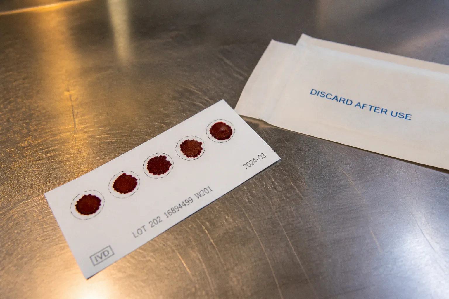
The heel-prick method uses capillary blood collected onto a filter paper card. The dried blood spot is transported to a public health or clinical laboratory, where a punch from the card is processed for DNA extraction and TREC quantification.
In many programs, collection occurs within 24 to 48 hours after birth. Some protocols allow collection up to approximately 5 days of age. The exact timing is determined by the newborn screening system, discharge practices, and local regulatory requirements.
The method has several structural advantages.
Population-scale throughput
Dried blood spot cards are compatible with centralized laboratory workflows. Multiple newborn screening assays can be performed from the same card or from coordinated specimens. The format supports automation, batch processing, barcode tracking, and standardized result reporting.
This matters because SCID screening is not a low-volume diagnostic service. A method may be technically valid but operationally unsuitable if it cannot maintain throughput, sample traceability, and short turnaround times across an entire birth cohort.
Established quality-control infrastructure
Heel-prick collection is integrated into newborn screening programs in many jurisdictions. Laboratories have defined procedures for:
- Card labeling and accessioning.
- Specimen drying and transport.
- DNA extraction.
- Internal amplification controls.
- Cutoff calibration.
- Repeat testing.
- Referral of abnormal results.
- Management of insufficient or invalid specimens.
The quality of the assay depends on the full chain, not only on PCR performance. Incomplete blood saturation, layering of drops, contamination, excessive humidity, delayed transport, and poor punching can affect DNA recovery and analytical interpretation.
Standardized public health implementation
TREC testing from dried blood spots remains the global population-wide standard for newborn SCID screening. It was piloted in Wisconsin in 2008, introduced in Ontario in 2013, and achieved nationwide implementation across all 50 US states in 2018.
The strength of this approach is not that it eliminates every false-positive result. Its strength is that it combines a clinically relevant biomarker with a scalable specimen format and an established referral pathway.
Limitations of the heel-prick specimen
The heel-prick sample is not collected immediately at birth. That delay is usually clinically acceptable because screening programs are designed around early postnatal collection and rapid follow-up. It can nevertheless create operational problems when:
- The infant is discharged before sampling.
- The specimen is collected too early or too late for the local protocol.
- The card is inadequately filled.
- The sample is lost or delayed in transit.
- The infant is receiving intensive neonatal care.
- A repeat specimen is required before referral.
The method also has a known limitation in premature infants. Developmental immaturity is associated with lower TREC levels and higher repeat-testing or false-positive rates. A low result in a premature infant therefore has to be interpreted in the context of gestational age, birth weight, clinical status, and specimen timing.
Umbilical cord blood TREC testing and flow cytometry
Umbilical cord blood provides a specimen at the moment of delivery. This removes the postnatal collection interval and avoids the need for a separate heel-prick procedure. Research and alternative clinical protocols have evaluated two main strategies:
1. TREC analysis performed on cord blood.
2. Flow cytometric evaluation of cord-blood lymphocyte populations.
These approaches should not be grouped as a single test. TREC analysis is a molecular assay for thymic output. Flow cytometry is a cellular assay that characterizes immune-cell subsets. Their analytical outputs, controls, and failure modes differ.
Cord blood has been reported to contain approximately 4.5 times higher baseline TREC counts per sample than adult peripheral blood. That higher signal may provide analytical headroom for TREC measurement. It does not, by itself, establish superior sensitivity or specificity compared with dried blood spot screening in a national newborn cohort.
Immediate specimen availability
The most direct advantage of cord blood is timing. The sample can be obtained immediately after birth, before routine newborn screening workflows begin. This could be relevant in settings where early identification changes the initial management pathway, including infection avoidance, immunoglobulin planning, and referral to a specialist center.
The practical value depends on processing capacity. A specimen collected at birth is not clinically useful if it remains unprocessed or if abnormal results cannot trigger confirmatory testing rapidly.
Flow cytometric information
Flow cytometry can measure the absolute or relative distribution of T cells, B cells, and natural killer cells. It can also characterize naïve T-cell populations and other immunophenotypic features relevant to inborn errors of immunity.
This provides more direct cellular information than TREC quantification alone. However, flow cytometry requires:
- Fresh or appropriately preserved cellular material.
- Rapid transport and processing.
- Validated antibody panels.
- Instrument calibration.
- Skilled technical interpretation.
- Reliable absolute lymphocyte counts.
- Age-appropriate reference intervals.
A cord-blood flow cytometry program is therefore more laboratory-intensive than a dried blood spot PCR workflow. It may be highly informative in a specialist setting, but population-scale deployment requires additional infrastructure.
Preanalytical variables
Cord blood collection has its own technical vulnerabilities. Sample quality can be affected by:
- Maternal blood contamination.
- Delayed clamping or collection procedures.
- Clot formation.
- Insufficient volume.
- Variable anticoagulant ratios.
- Delayed delivery to the laboratory.
- Perinatal stress and changes in leukocyte distribution.
These variables do not invalidate cord blood as a specimen. They define the quality-control requirements. A cord-blood assay must demonstrate that the collection process produces a stable, representative specimen across routine deliveries rather than only under controlled research conditions.
Cord blood versus heel prick: method comparison
The comparison below separates operational characteristics from diagnostic claims. Evidence for cord blood approaches remains less standardized than evidence for heel-prick dried blood spot screening.
| Parameter | Heel-prick dried blood spot | Umbilical cord blood |
|---|---|---|
| Collection time | Usually 24–48 hours after birth; some protocols allow collection up to 5 days | Immediately at delivery |
| Primary SCID biomarker | TREC quantification from dried blood spot DNA | TREC quantification or lymphocyte immunophenotyping by flow cytometry |
| Main analytical output | Indirect estimate of thymic T-cell output | Thymic-output estimate or direct cellular phenotype, depending on assay |
| Laboratory workflow | Highly compatible with centralized, automated newborn screening | Requires validated molecular or cellular workflow; flow cytometry requires fresh-cell handling |
| Throughput | High and established for population screening | Potentially scalable, but workflow requirements vary substantially |
| Transport stability | Dried specimen is relatively suitable for centralized transport | Cellular assays are more sensitive to transport and processing delays |
| False-positive pressure | Increased in premature infants and with poor specimen quality | Potential effects from gestational age, collection quality, cellular composition, and reference intervals |
| Confirmatory testing | Required after abnormal TREC result | Required after abnormal TREC or flow cytometry result |
| Current status | Established population-wide standard | Investigated alternative; not a replacement global standard |
| Main advantage | Standardization and public health scalability | Immediate sampling and possible earlier access to cellular data |
| Main limitation | Collection occurs after birth and may require repeat sampling | Greater preanalytical and laboratory complexity, especially for flow cytometry |
The table also illustrates why the phrase “heel prick test versus cord blood” can be misleading. There is no single cord-blood assay equivalent to the dried blood spot TREC test. The comparison depends on whether the cord specimen is analyzed by PCR-based TREC quantification, flow cytometry, or a combined algorithm.
Diagnostic accuracy is determined by the pathway, not the specimen alone
Sensitivity and specificity cannot be assigned to a specimen type without defining the assay, cutoff, population, and reference standard. A TREC assay may have excellent analytical sensitivity for its molecular target while still producing a clinically non-specific result because low TRECs occur in conditions other than SCID.
The principal diagnostic metrics require separate interpretation:
- Analytical sensitivity: the ability of the assay to detect low levels of TREC target material.
- Clinical sensitivity: the ability to identify infants who truly have the target immune disorder.
- Specificity: the ability to classify infants without the disorder as screening-negative.
- Positive predictive value: the probability that an abnormal result represents clinically significant disease in the screened population.
- Invalid or insufficient specimen rate: the proportion of samples that cannot be interpreted reliably.
Cord blood may offer a higher baseline TREC signal than adult blood. That finding is relevant to assay design but is not equivalent to proof of higher clinical sensitivity. A high signal can improve signal separation in some platforms. It can also interact with cell composition, gestational age, and cutoff selection.
Similarly, flow cytometry can provide direct lymphocyte counts but does not automatically provide better population-screening specificity. Reference intervals must account for gestational age and the normal transition in neonatal immune-cell populations. A test that is biologically detailed can still be operationally inefficient if it generates excessive referrals from borderline findings.
The role of confirmatory testing
An abnormal screening result should lead to a controlled diagnostic sequence rather than immediate disease labeling. Confirmatory flow cytometry is central because it determines whether the infant has significant T-cell lymphopenia and defines the broader lymphocyte phenotype.
Genetic sequencing is then used to investigate pathogenic variants. Depending on the clinical and laboratory pattern, the testing strategy may include a targeted panel for known immune-deficiency genes, broader exome sequencing, or another validated sequencing approach. The selection depends on phenotype, family history, prior results, laboratory resources, and turnaround requirements.
A negative genetic result does not automatically negate an abnormal immune phenotype. Conversely, the identification of a variant requires classification against established evidence. A sequence change is not a diagnosis merely because it appears in a gene associated with immunodeficiency.
The newborn SCID screening timeline
The practical value of either specimen depends on how quickly the result moves through the clinical system. A functional newborn SCID screening timeline contains several stages:
1. Specimen collection. Heel-prick collection usually occurs at 24–48 hours after birth or according to the local protocol. Cord blood can be collected at delivery.
2. Primary assay. TREC analysis is performed on the dried blood spot or cord specimen. Cord blood may alternatively undergo flow cytometric assessment.
3. Result classification. The laboratory reports a screen-negative, abnormal, or invalid result according to validated thresholds.
4. Immediate risk management. An infant with a significantly abnormal result may require protective measures while confirmation is pending, based on specialist guidance.
5. Confirmatory flow cytometry. T-cell, B-cell, and natural killer cell populations are quantified. Naïve T-cell status may be included.
6. Molecular investigation. Genetic testing evaluates pathogenic variants associated with the phenotype.
7. Definitive clinical pathway. The infant is referred for immune-deficiency management, infection prevention, and consideration of HSCT where indicated.
The difference between collection at birth and collection at 24–48 hours is clinically relevant only if each downstream stage is equally efficient. A cord-blood method can reduce the time to specimen acquisition. It cannot compensate for delayed laboratory processing, slow referral, or unavailable confirmatory testing.
The shortest collection interval is not the same as the shortest diagnostic interval.
For infants with confirmed SCID, early intervention is the relevant endpoint. HSCT before 3.5 months of age is associated with significantly better survival and outcomes than later treatment. Screening policy should therefore be judged by the complete time from birth to confirmed diagnosis and treatment eligibility, not by specimen collection time in isolation.
Premature infants and low TREC results
Prematurity is a major confounder in newborn TREC screening. The thymus and peripheral immune system are developmentally immature, and premature infants may show lower TREC levels without having SCID. The result is an increased rate of abnormal or repeat screens.
This is a specificity problem, not a reason to exclude premature infants from screening. A premature infant with a low TREC count still requires a defined follow-up pathway because true T-cell lymphopenia can occur in this population.
Interpretation should incorporate:
- Gestational age at birth.
- Birth weight.
- Postnatal age at collection.
- Current clinical condition.
- Transfusions or other interventions.
- Whether the specimen is a first screen or a repeat.
- The magnitude of the TREC reduction.
- Results from confirmatory flow cytometry.
The same cutoff may not perform identically across term and premature populations. Programs may use repeat testing, adjusted thresholds, or direct referral for flow cytometry in selected high-risk groups. The chosen algorithm should minimize missed disease without creating an unmanageable burden of unnecessary confirmatory testing.
Cord blood does not remove this biological issue. The specimen is collected earlier, but developmental immune immaturity remains present. Cord-blood reference intervals and cutoff performance must therefore be validated across gestational ages and delivery contexts.
Where cord blood may provide a practical advantage
Cord blood screening has a plausible operational role in settings that already possess delivery-room collection protocols and rapid immunology laboratory access. Its strongest potential advantages are specific:
- Sampling occurs before postnatal discharge.
- The specimen is available before the standard heel-prick collection window.
- Flow cytometry can provide direct information on lymphocyte subsets.
- Cord blood may show a higher baseline TREC signal than adult peripheral blood.
- The initial immune assessment could be integrated into specialist neonatal pathways.
These advantages are conditional. Cord-blood screening requires a reliable chain from obstetric collection to laboratory accessioning. It also requires clear handling of samples that are clotted, contaminated, insufficient, or delayed.
The method is more likely to be useful as an institutional or regional protocol than as a universal replacement unless large-scale performance data establish comparable or superior sensitivity, specificity, invalid rates, cost, and clinical outcomes.
Where heel-prick screening remains stronger
Heel-prick dried blood spot screening remains the more mature public health method because its laboratory and governance systems are already established. Its advantages include:
- High-throughput processing.
- Stable specimen format.
- Existing newborn screening logistics.
- Broad experience with cutoff management.
- Standardized referral pathways.
- Compatibility with centralized testing laboratories.
- Lower dependence on immediate cellular viability.
The method is not analytically perfect. It is operationally optimized. In population screening, that distinction is decisive.
A cord-blood flow cytometry model may produce richer biological data per infant but require more personnel, more rapid specimen handling, and more specialized instrumentation. A dried blood spot TREC assay may produce a narrower output but achieve better consistency across thousands of samples.
Clinical utility: what should determine method selection
The selection between cord blood and heel prick should be based on clinical utility rather than on the apparent sophistication of the assay. A valid implementation must answer five technical questions:
- Does the method detect clinically significant T-cell lymphopenia with adequate sensitivity?
- Does it maintain acceptable specificity in premature and medically complex infants?
- Can the laboratory process specimens at the required throughput?
- Can abnormal results reach an immunology service without delay?
- Does the pathway improve time to confirmed diagnosis and definitive treatment?
The current evidence supports a clear distinction. Heel-prick dried blood spot TREC screening is the established standard for population-wide SCID screening. Umbilical cord blood is a credible alternative specimen under investigation, with potential advantages in immediate collection and cellular assessment. However, available evidence does not establish cord blood flow cytometry or cord-blood TREC testing as a universal replacement.
A technically defensible diagnostic pathway uses the methods in sequence:
1. Population screening with a validated TREC assay.
2. Rapid confirmation with flow cytometry.
3. Molecular testing for pathogenic variants when indicated.
4. Specialist management based on the integrated immune phenotype.
The specimen is only the first variable. The decisive performance metric is the interval from birth to a clinically actionable diagnosis. Until cord-blood protocols demonstrate equivalent or superior performance across large populations, the heel-prick dried blood spot remains the more reliable population-screening platform, while cord blood retains value as an early-sampling strategy and a subject of targeted clinical research.