JIA uveitis screening: how often does your child need it?
In my line of work, I see clinical protocols that are meticulously designed and then routinely weakened at the point of care. JIA uveitis screening sits high on that list.
The prevalence numbers are stark: uveitis affects roughly 10% to 30% of children with juvenile idiopathic arthritis overall, rising to about 45% to 57% in some young, ANA-positive oligoarticular groups. More importantly, it is often asymptomatic until inflammation has already caused measurable damage.
The 2019 ACR/Arthritis Foundation guideline and the 2023 Canadian Rheumatology Association consensus both describe a risk-based screening approach. Yet pediatric rheumatology charts still arrive where a slit-lamp examination is treated as optional, or where a standard school vision screening is assumed to have covered the relevant risk. It has not.
The practical question is not simply whether a child has a positive ANA. The question is how ANA status fits with the JIA subtype, the child’s age when arthritis began, and how long the child has had JIA. Those factors work together to determine the screening frequency.
The Silent Threat: Why Standard Eye Exams Miss JIA Uveitis
Here is the uncomfortable truth that gets glossed over in patient handouts: JIA-associated chronic anterior uveitis is usually a silent disease. There may be no pain, no obvious redness, no light sensitivity, and no complaint that a parent can identify across the breakfast table. A child may continue reading, attending school, and playing normally while inflammation is developing in the anterior chamber.
By the time blurred vision becomes noticeable, structural complications may already be present. These can include synechiae, band keratopathy, cataract, macular edema, or elevated intraocular pressure. The point of screening is to find inflammation before the child has a reason to report it.
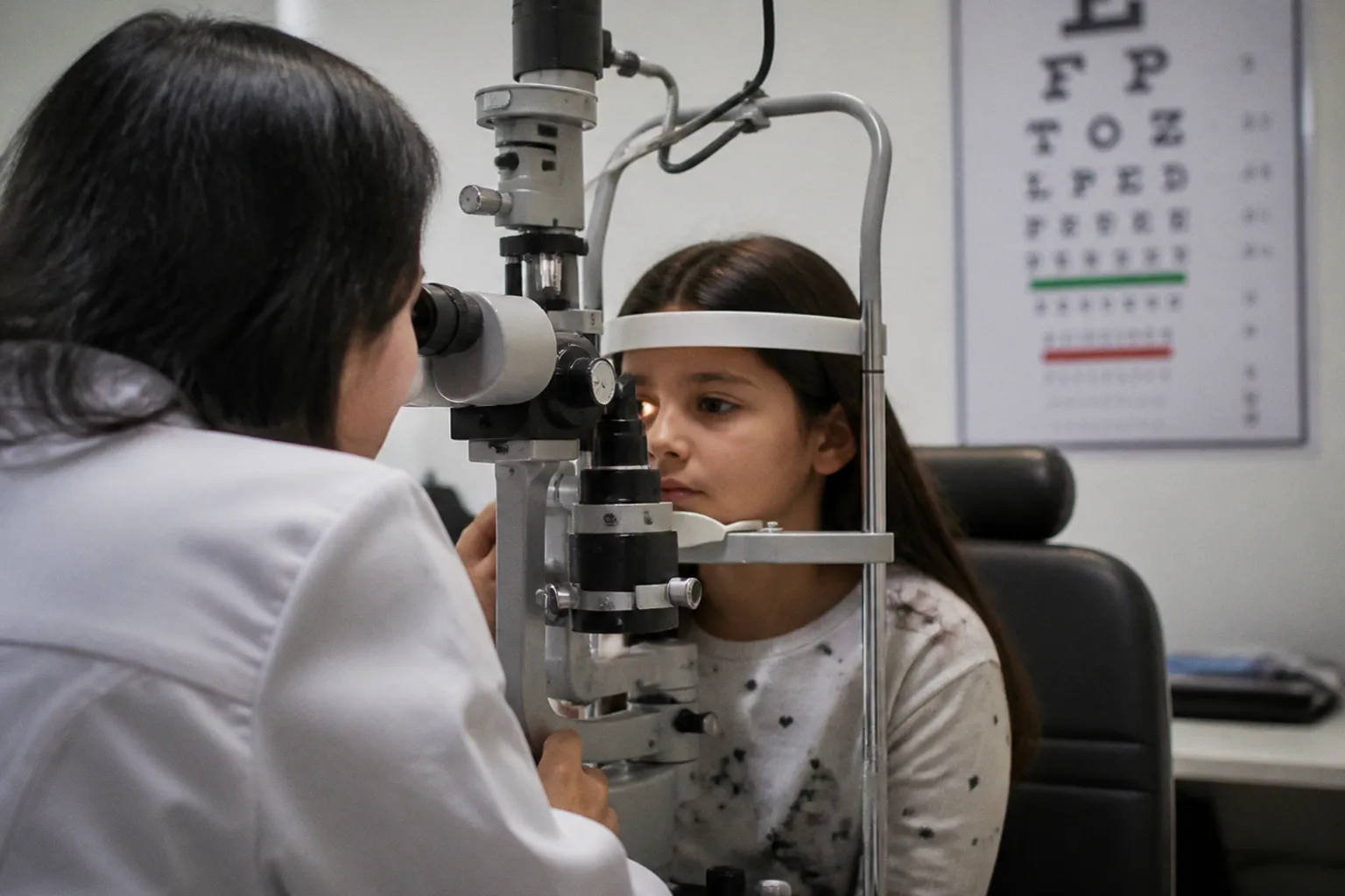
That is why the slit-lamp examination is central. A direct ophthalmoscope, a penlight examination, a school nurse’s vision chart, or a routine refraction may be useful for other purposes, but none of them reliably replaces a slit-lamp assessment for anterior uveitis. The slit lamp allows the ophthalmologist to examine the anterior chamber and grade cells and flare—the findings on which the screening and treatment decisions are based.
The distinction is easy to lose in a referral pathway. “Eye exam” can mean a number of different things. For a child with JIA, the referral should make clear that the purpose is uveitis screening and that a slit-lamp examination is required. The examination should be performed by an ophthalmologist or by an appropriately trained eye-care professional working within the relevant ophthalmology pathway.
A normal vision chart is reassuring about visual acuity. It is not proof that a child with JIA has no uveitis.
What repeatedly causes trouble is not always a lack of medical knowledge. It is the handoff. A family hears that the child needs an eye exam, schedules with the nearest available provider, and receives a standard refractive assessment. The child is told that vision is normal. Everyone leaves with the impression that screening has been completed, although the specific examination needed to detect anterior chamber inflammation never took place.
That is the failure mode I would eliminate first from patient-facing instructions: replacing a defined ophthalmic examination with the vague phrase “routine eye check.”
Risk Stratification: How ANA Status and Age Dictate Screening Intervals
The screening cadence is not arbitrary, but it is also not determined by one laboratory result. A positive antinuclear antibody test is an important marker of increased uveitis risk in JIA. It is not a stand-alone diagnosis, and it does not override the rest of the clinical picture.
The risk assessment should bring together at least four elements:
1. ANA status. ANA positivity is associated with a higher risk of chronic anterior uveitis, particularly in younger children with certain JIA patterns. ANA negativity lowers risk but does not eliminate it. A child with ANA-negative JIA can still develop uveitis and still needs screening.
2. Age at arthritis onset. Younger age at onset is associated with greater uveitis risk than onset in adolescence. Age matters partly because it interacts with the disease phenotype and ANA status. A young child with ANA-positive oligoarticular JIA does not have the same risk profile as an older child with a different subtype and the same laboratory result.
3. JIA subtype. Oligoarticular JIA and rheumatoid factor-negative polyarticular JIA are commonly included among the higher-risk categories. Systemic JIA is generally associated with a lower risk of chronic anterior uveitis, while enthesitis-related and psoriatic forms require interpretation in the context of their own clinical features and the child’s other risk markers.
4. Disease duration. Risk is often greatest during the first several years after JIA begins. That is why screening is usually more frequent early in the disease course and may be extended later when a child has remained free of uveitis.
ANA status is therefore one input into the risk algorithm, not the algorithm itself. The clinically useful statement is not that a positive ANA automatically places every child in the highest-risk tier. It is that ANA positivity can move a child toward a higher-risk schedule when it appears alongside the relevant age, subtype, and disease-duration factors.
This distinction matters in both directions. Overstating the role of ANA can lead families to believe that a positive result determines everything. Understating it can lead to a high-risk child being screened too infrequently. The proper interpretation is combined risk assessment.
Why the ANA result needs context
ANA testing is not a simple “safe” or “unsafe” switch. The result is interpreted alongside the child’s clinical diagnosis and, where relevant, the laboratory method and reporting convention used by the testing laboratory. A positive ANA does not predict that uveitis will definitely develop. It identifies a pattern associated with increased probability in a particular JIA context.
Nor does an ANA-negative result make symptoms irrelevant. If a child develops eye pain, marked light sensitivity, redness, a new visual complaint, or any other concerning change, that is no longer a routine screening question. The child should be assessed promptly, regardless of the previous ANA result or the date of the last scheduled examination.
The same principle applies to disease activity. Remission of joint symptoms is clinically important, but it does not automatically erase the child’s historical uveitis risk. A child may need ongoing screening even when the arthritis is well controlled, including when control depends on medication.
The 3–6–12 Month Rule: Understanding Your Child’s Specific Schedule
In its familiar form, the screening framework divides children into broad risk tiers. The intervals below are useful shorthand, but they are not a substitute for the treating team’s assessment. Guidelines and local pathways may differ in how they define the categories, especially for children who do not fit neatly into one group.
| Broad risk profile | Factors that may contribute | Typical screening interval |
|---|---|---|
| Higher risk | Often includes younger age at onset, ANA positivity, oligoarticular or rheumatoid factor-negative polyarticular JIA, and the early years after diagnosis | Every 3 months |
| Intermediate risk | A mixed profile, such as one or more risk factors without the full higher-risk combination | Every 6 months |
| Lower risk | Often includes older age at onset, lower-risk JIA patterns, ANA negativity, or a longer period without uveitis | Every 12 months |
The table should not be read as a scoring system in which one positive feature automatically decides the interval. A child with ANA positivity but an older age at onset and a lower-risk JIA subtype may not have the same schedule as a young child with ANA-positive oligoarticular disease. Conversely, a child without ANA positivity may still require more than annual screening if other clinical considerations raise concern.
The 3-month interval is used for children whose combined profile places them at higher risk. The 6-month interval is used for children in an intermediate group, where annual screening may be too widely spaced but quarterly examinations may not be necessary. The 12-month interval is generally reserved for children whose overall risk is lower, not for every child who happens to be ANA-negative.
The schedule also assumes that the child is being screened for asymptomatic disease. Once active uveitis is identified, the situation changes. Follow-up is then determined by the severity of inflammation, complications, treatment response, and the ophthalmologist’s management plan. The screening calendar no longer governs care.
In practice, the intermediate group is where scheduling becomes least reliable. These children do not always look “high risk” at a glance, so a six-month examination can quietly become an annual appointment. That is not a harmless administrative drift. The interval should be chosen deliberately from the child’s combined risk profile and recorded in a way that the family, rheumatology team, and ophthalmology team can all see.
The schedule should be a documented clinical decision, not a default interval that survives because nobody changed it.
The Critical First Window: Timing the Initial Post-Diagnosis Exam
The first slit-lamp examination is not simply the first item in the 3–6–12 cycle. It is a separate baseline assessment and should be arranged as soon as reasonably possible after the JIA diagnosis is established.
The ACR/Arthritis Foundation guideline places initial screening within the first one to three months after diagnosis. Earlier standards, including BSPAR guidance, have used a tighter window for children considered at risk. Local access and the child’s clinical circumstances can affect the exact pathway, but the referral should begin at diagnosis rather than being left until a later rheumatology review.
The reason is straightforward: uveitis may already be present when arthritis is diagnosed. A baseline examination documents the condition of the eyes at the start of the child’s rheumatology care and gives the ophthalmologist a reference point for later findings. Without that baseline, it becomes harder to determine whether inflammation at a subsequent appointment is new, persistent, or simply newly detected.
The referral itself should carry useful information. It should identify:
- the child’s JIA diagnosis and subtype, if established;
- age at arthritis onset;
- ANA status, interpreted in the wider clinical context;
- the date of diagnosis;
- the reason for referral—JIA-associated uveitis screening;
- the need for a slit-lamp examination rather than a vision check alone.
A generic referral asking an eye-care provider to “evaluate” the child leaves too much room for the wrong kind of appointment. Families should not have to guess whether the scheduled visit includes the examination required for JIA uveitis screening.
The first appointment can be delayed by insurance authorization, limited pediatric ophthalmology capacity, transport problems, or the ordinary shock of receiving a chronic-disease diagnosis. These are real barriers, not failures of commitment. They are also reasons to start the referral process immediately and to ask the rheumatology team what to do if the recommended appointment cannot be obtained within the intended window.
If the child develops eye symptoms while waiting, the family should not wait for the routine screening appointment. Pain, redness, light sensitivity, a change in vision, or a child suddenly holding reading material unusually close all warrant clinical advice. Although JIA uveitis is often silent, symptoms should never be dismissed simply because the child is already on a screening pathway.
Navigating Long-Term Monitoring: When Screening Frequency Changes
The screening schedule should evolve with the child’s risk profile, but changes should be explicit. A calendar that was appropriate at diagnosis may no longer be appropriate several years later, and an old interval should not remain in place merely because it is embedded in the electronic record.
The most common planned transition is a step-down from three-monthly screening after a child has moved beyond the period of greatest risk without developing uveitis. The precise timing depends on the child’s age, JIA subtype, ANA status, disease duration, and the guideline used by the treating team. Passing a particular anniversary is not, by itself, a reason to change the interval. The change should follow a review of the entire risk profile.
There can also be reasons to increase attention rather than reduce it. A change in the working diagnosis or JIA subtype may alter the risk assessment. New systemic or musculoskeletal features may prompt the team to reconsider whether the original category still fits. A flare of arthritis does not automatically mean that the child has active uveitis, but it is an opportunity to confirm that ophthalmology follow-up is current.
ANA results require particular care here. ANA status is not usually a marker that should be treated as a constantly changing traffic signal. Repeat testing may occur in clinical practice, but a later positive or negative result should not be used in isolation to redesign the child’s screening schedule. The meaningful assessment remains the combination of the child’s JIA phenotype, age at onset, disease course, and other clinical factors.
The third important transition is from screening to monitoring known uveitis. Once inflammation has been diagnosed, examinations may be needed substantially more often than every three, six, or twelve months. The ophthalmologist may shorten the interval during treatment initiation or adjustment and extend it only when the inflammation is controlled and the treatment plan allows. A child with active disease should not be managed according to a routine screening calendar.
Practical signals that the schedule needs review
Several patterns should prompt a family to ask the rheumatology team for clarification:
- The child’s only recent “eye exam” was a school vision screening, a vision-chart check, or a routine refraction, with no clear documentation of a slit-lamp examination.
- The chart does not state the child’s current risk profile or the next recommended screening interval.
- The family has been told that screening can stop because the joints are quiet or the child is in remission on treatment, without a documented ophthalmology decision.
- The child has moved from the early years after diagnosis into a later stage of disease, but nobody has discussed whether the interval should remain the same.
- The child’s JIA diagnosis or subtype has changed, but the ophthalmology schedule was never reconsidered.
- The family is unsure whether the ophthalmologist knows that the visit is for JIA uveitis screening.
These are not accusations of negligence. They are common system-level failure points: unclear referrals, disconnected records, and schedules that are never revisited. Naming the problem gives the family and clinical team something concrete to fix.
What parents should ask at the visit
The most useful questions are specific:
1. What is my child’s current uveitis-risk profile?
2. How were ANA status, age at onset, JIA subtype, and disease duration considered together?
3. Does the next appointment include a slit-lamp examination?
4. How often should screening occur, and when should that interval be reviewed?
5. Who should the family contact if eye symptoms appear between scheduled visits?
6. If the child’s joints are controlled, does the ophthalmology schedule change—and who is making that decision?
A clear answer should produce a clear date or interval. “We will monitor” is not a schedule.
The point is precision, not panic
JIA uveitis screening is a high-value part of pediatric rheumatology because it is designed for a disease that often gives families no early warning. The screening method matters: a standard vision check is not the same as a slit-lamp examination. The timing matters: the initial examination establishes a baseline, and the early years after diagnosis generally require closer attention. The interpretation matters most of all: ANA status is important, but it must be read alongside age at onset, JIA subtype, and disease duration.
For most families, the practical priorities are straightforward. Confirm that the child has had the correct examination, make sure the current risk profile is documented, and ask for a specific screening interval rather than a vague plan to keep watching. The familiar 3–6–12 month framework is useful, but it is only the framework. The actual schedule belongs to the child in front of the clinician—not to the ANA result alone.