Thymus transplant vs stem cell transplant for athymia
Reported overall survival after hematopoietic stem cell transplantation in congenital athymia is approximately 41%, compared with survival approaching 90% in typical severe combined immunodeficiency caused by hematopoietic stem cell defects.
The difference is mechanistic, not procedural. HSCT replaces defective blood-forming cells. It does not create a functional thymic epithelium.
Congenital athymia is therefore not a standard SCID transplant problem. The central abnormality is the absence or severe failure of the thymus, the organ required for de novo T-cell maturation and selection. In complete DiGeorge syndrome, CHARGE syndrome, or rare FOXN1 and PAX1 defects, donor stem cells may engraft without producing a durable, diverse naïve T-cell compartment.
The relevant comparison is not simply transplant versus transplant. It is thymic tissue replacement versus hematopoietic replacement.
The biological defect determines the treatment route
T cells do not become fully functional merely because hematopoietic stem cells are present. Precursor T cells must migrate to a functioning thymus. Inside the thymic environment, they undergo proliferation, receptor rearrangement, positive selection, negative selection, and maturation into naïve T cells with a broad T-cell receptor repertoire.
This sequence fails in congenital athymia.
The condition is most strongly associated with complete DiGeorge syndrome, frequently linked to a 22q11.2 deletion. It can also occur in CHARGE syndrome and in patients with pathogenic variants affecting FOXN1 or PAX1. These disorders differ genetically, but the diagnostic endpoint is similar: profound failure of thymic function and inadequate production of naïve T cells.
The distinction from typical SCID is operationally important:
- In hematopoietic SCID, the primary defect is within the blood-forming or lymphoid compartment. HSCT can supply donor-derived progenitors capable of generating T cells.
- In congenital athymia, the hematopoietic precursors may be present or replaceable, but the stromal organ required for T-cell maturation is absent or nonfunctional.
- Donor mature T cells transferred with an HSCT graft can provide temporary immune activity. This is not equivalent to native thymopoiesis.
- A high donor T-cell count after HSCT does not necessarily indicate restoration of a diverse, self-tolerant naïve T-cell pool.
This is why the phrase “stem cell transplant for athymia” requires qualification. HSCT may be used as a rescue or alternative strategy, but it does not correct the intrinsic thymic defect.
The transplant target must match the defective compartment: hematopoietic stem cells for hematopoietic failure, thymic tissue for thymic failure.
Thymus tissue transplantation: replacing the missing maturation site
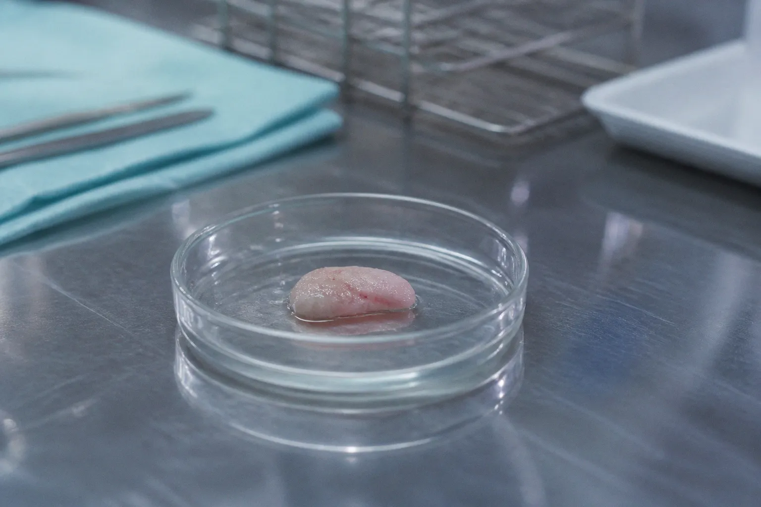
Allogeneic thymus transplantation uses processed postnatal thymic tissue. The tissue is generally obtained from infants undergoing cardiothoracic surgery and prepared before implantation. It is placed into the recipient, commonly within the quadriceps muscle, where it can support the development of recipient-derived T cells.
The donor tissue is not intended to supply a permanent population of donor lymphocytes. Its clinical function is different. It provides a thymic epithelial microenvironment in which the recipient’s hematopoietic precursors can undergo T-cell maturation.
Over time, the expected biological sequence is:
1. Recipient hematopoietic precursors enter the implanted thymic tissue.
2. T-cell receptor gene rearrangement produces a diverse repertoire.
3. Developing T cells undergo selection within the donor thymic epithelium.
4. Naïve T cells enter peripheral blood.
5. T-cell diversity and immune competence expand progressively rather than appearing immediately after implantation.
This time course has direct implications for infection control. Thymus transplantation does not provide immediate immune reconstitution equivalent to passive donor lymphocyte transfer. The child may remain highly vulnerable during the period required for thymopoiesis.
Allogeneic processed thymus tissue, including products such as processed thymus tissue-agdc, is therefore a specialized therapy for a narrow phenotype. It is not a general treatment for every child with low T-cell numbers, 22q11.2 deletion, or recurrent infection.
Diagnostic thresholds before thymus transplantation
Confirmation of congenital athymia depends on cellular immunophenotyping. Flow cytometry is central. A commonly used diagnostic threshold is:
- fewer than 50 naïve T cells per mm³ in peripheral blood, or
- fewer than 5% of total T cells displaying a naïve phenotype.
The phenotype must be interpreted with age, clinical history, infection status, prior transfusions, and treatment exposure. A single low lymphocyte count is insufficient. Total T-cell numbers can be misleading because circulating mature or activated T cells may be present despite absent thymic output.
The laboratory assessment should distinguish:
- total CD3-positive T cells;
- CD4 and CD8 subsets;
- naïve versus memory phenotype;
- recent thymic emigrant markers where available;
- T-cell receptor repertoire diversity;
- proliferative responses to mitogens and antigens;
- maternal engraftment or transfusion-associated lymphocytes when clinically relevant.
T-cell receptor excision circles can support evidence of thymic output in screening and follow-up, but they do not replace a complete immunophenotyping strategy. In a child with suspected complete DiGeorge syndrome, the assay must answer a specific question: is there meaningful endogenous thymopoiesis, or is the thymic compartment functionally absent?
HSCT: a different graft for a different failure mode
HSCT is established therapy for many forms of severe combined immunodeficiency. In those disorders, donor hematopoietic stem cells can repopulate the marrow and generate lymphoid lineages. The expected result is donor-derived immune reconstitution, provided that engraftment, infection control, conditioning, and graft-versus-host disease management are adequate.
Congenital athymia does not provide the same biological substrate.
Following HSCT, mature donor T cells may circulate and temporarily improve immune surveillance. However, without a functional thymus, the graft may not generate a stable supply of newly educated naïve T cells. The repertoire can remain restricted. Peripheral T-cell expansion can be oligoclonal. Immune function may depend on the survival and proliferation of cells transferred in the graft rather than on continuous de novo production.
This creates several technical problems:
- T-cell numbers may increase without normalization of repertoire diversity.
- Donor mature T cells can recognize host tissues and produce GVHD.
- Immunity may deteriorate as transferred T cells contract.
- The graft does not repair a FOXN1-, PAX1-, or 22q11.2-related thymic stromal defect.
- Conditioning toxicity and infectious complications remain relevant even when long-term thymopoiesis is inadequate.
Reported GVHD incidence in HSCT-treated congenital athymia is approximately 50% to 56%. A Japanese nationwide retrospective study covering 2000–2024 reported a one-year overall survival of 66.7% for allogeneic HCT used as a rescue strategy. These data support HSCT as a possible option in selected circumstances, but they do not establish equivalence with thymus transplantation.
The comparison is also affected by case selection. Children treated with rescue HSCT may have severe infection, organ damage, lack of access to thymus tissue, or delayed diagnosis. The observed survival rate is therefore not a direct head-to-head estimate of procedure quality. It is a clinically relevant signal of risk in a difficult population.
Direct comparison of the two approaches
| Parameter | Thymus tissue transplantation | Hematopoietic stem cell transplantation |
|---|---|---|
| Primary target | Defective or absent thymic stromal environment | Defective hematopoietic and lymphoid compartment |
| Mechanism | Provides tissue for recipient T-cell maturation and selection | Provides donor hematopoietic stem cells and, depending on graft, mature donor lymphocytes |
| Corrects absent thymic epithelium | Yes, as the intended mechanism | No |
| De novo naïve T-cell production | Expected after tissue engraftment and maturation | Limited or absent when intrinsic athymia persists |
| T-cell repertoire | Intended to develop through thymic selection | May remain restricted if driven mainly by transferred mature T cells |
| Immediate immune effect | Delayed; thymopoiesis requires time | Can be faster if mature donor T cells are present |
| GVHD risk | Generally not driven by a conventional marrow graft | Major risk, particularly when mature donor T cells are involved |
| Typical clinical role | Definitive treatment for confirmed congenital athymia | Rescue or alternative treatment when thymus transplantation is unavailable or unsuitable |
| Key eligibility signal | Profound naïve T-cell deficiency confirmed by flow cytometry | A clinical decision based on disease severity, donor options, and lack of a functioning thymus |
| Main limitation | Specialized availability and delayed immune recovery | Does not restore thymic architecture and may produce GVHD without durable thymopoiesis |
This table should not be read as a simplified superiority ranking. The procedures address different anatomical and cellular defects. The decisive variable is whether the child has a transplantable thymic target and whether the diagnosis confirms true athymia rather than a less severe thymic hypoplasia.
Complete DiGeorge syndrome: treatment selection is phenotype-dependent
Complete DiGeorge syndrome is the most recognizable setting for congenital athymia. The 22q11.2 deletion itself does not determine treatment. The degree of thymic dysfunction does.
Many children with 22q11.2 deletion have partial thymic hypoplasia rather than complete athymia. They may produce naïve T cells, although counts can be reduced and immune responses may be variable. These children are not automatically candidates for thymus tissue transplantation or HSCT.
The relevant assessment includes:
- absolute naïve CD4 and CD8 T-cell counts;
- evidence of recent thymic emigrants;
- mitogen and antigen responses;
- infection burden;
- vaccine response interpretation;
- cytopenias and platelet abnormalities;
- calcium and parathyroid function;
- cardiac anatomy and surgical history;
- evidence of autoimmunity;
- genetic confirmation and phenotype correlation.
In complete DiGeorge syndrome, the combination of profound naïve T-cell deficiency and absent functional thymic output establishes a fundamentally different risk category. Opportunistic infections can become fatal within the first two to three years without therapeutic intervention or an effective salvage strategy.
The treatment route must also account for the associated phenotype. Children with complete DiGeorge syndrome may have congenital heart disease, hypocalcemia, airway abnormalities, feeding problems, developmental complications, and autoimmune manifestations. These factors affect anesthesia, conditioning, infection management, nutritional support, and post-transplant surveillance.
A normal or near-normal total lymphocyte count does not exclude severe thymic dysfunction. The assay must be phenotype-resolved.
Partial thymic function changes the interpretation
Residual thymic activity can produce a clinically meaningful naïve T-cell population. In that setting, the management may involve infection prophylaxis, immunoglobulin replacement when indicated, selective vaccine restrictions, and longitudinal immune monitoring rather than immediate transplantation.
The distinction is not cosmetic. Thymus transplantation is an intervention for absent thymic function. It is not a treatment for every quantitative T-cell abnormality associated with a chromosomal deletion.
The clinical workflow is an assay and eligibility problem
A reliable pathway begins before a transplant team selects a graft. The first requirement is diagnostic separation among congenital athymia, typical SCID, combined immunodeficiency, and partial DiGeorge-associated immune dysfunction.
1. Establish the cellular phenotype
Flow cytometry should quantify total and naïve T-cell populations. The threshold of fewer than 50 naïve T cells/mm³ or fewer than 5% naïve T cells is a critical indicator used in evaluating congenital athymia for thymus tissue transplantation.
The report should not provide only percentages. Absolute counts are necessary. A low percentage can coexist with a clinically relevant absolute population, while a high percentage can be misleading when the total T-cell count is extremely low.
2. Exclude confounders
The laboratory interpretation must account for:
- recent blood product exposure;
- maternal T-cell engraftment;
- severe viral or fungal infection;
- immunosuppressive medications;
- prior chemotherapy or conditioning;
- age-dependent reference ranges;
- lymphocyte redistribution during acute illness.
These factors can distort the apparent T-cell phenotype. Repeat testing may be necessary when the clinical state has stabilized.
3. Define the genetic and syndromic diagnosis
Chromosomal microarray or a targeted 22q11.2 assay may identify the common deletion associated with DiGeorge syndrome. Broader sequencing can be required when the phenotype suggests CHARGE syndrome, FOXN1 deficiency, PAX1-related disease, or another inborn error of immunity.
A genetic result is not a substitute for functional immune profiling. Conversely, a negative targeted panel does not exclude a thymic developmental disorder if the assay lacks coverage of the relevant genes or structural variants.
The molecular report should specify:
- the gene and variant;
- zygosity;
- inheritance model;
- classification of pathogenicity;
- assay coverage and limitations;
- correlation with the cellular phenotype.
The term pathogenic variant should be reserved for variants meeting accepted classification standards. A variant of uncertain significance cannot independently establish the indication for transplantation.
4. Assess the infection and organ baseline
Both procedures become more hazardous in the presence of uncontrolled infection or end-organ injury. Baseline assessment commonly includes microbiological testing, imaging where indicated, hepatic and renal function, pulmonary evaluation, cardiac assessment, nutritional status, and immunoglobulin measurement.
The objective is not to generate a generic preoperative checklist. It is to quantify the factors that alter transplant risk and the expected value of immune reconstitution.
5. Select the route according to mechanism and availability
For confirmed congenital athymia, allogeneic thymus transplantation is the mechanism-matched definitive treatment when available and clinically feasible. HSCT may be considered when thymic tissue is unavailable, when a rescue strategy is required, or when the clinical team determines that the expected benefit outweighs the substantial risk of GVHD and incomplete immune restoration.
No single laboratory biomarker independently resolves this decision. The interpretation requires a concordant phenotype, genetic context, infection profile, and procedure-specific risk assessment.
An increase in circulating T cells is not equivalent to thymic reconstitution. The decisive endpoints are naïve-cell output, repertoire quality, immune function, and durability.
Risks after thymus transplantation
Thymus transplantation is specialized, but it is not low-risk. The implanted tissue requires time to support T-cell development. During this interval, infection prophylaxis and protective care remain necessary.
Potential complications include:
- persistent or recurrent opportunistic infection before immune recovery;
- inadequate thymic function after implantation;
- immune dysregulation;
- autoimmune complications;
- graft-related issues at the implantation site;
- complications associated with the underlying congenital syndrome;
- prolonged need for laboratory surveillance and supportive therapy.
The endpoint is not merely survival to discharge. A clinically useful outcome includes durable naïve T-cell production, functional immune responses, a sufficiently diverse receptor repertoire, and reduced dependence on replacement therapies.
Long-term surveillance is therefore required. Serial flow cytometry should evaluate the trajectory of naïve T-cell recovery rather than a single post-treatment value. T-cell receptor repertoire analysis may provide additional evidence of diversity where available. Functional assays remain relevant because phenotype and function are not interchangeable.
Risks after HSCT in congenital athymia
HSCT carries the standard hazards of allogeneic transplantation, with additional biological limitations in athymic patients.
The principal risks include:
- graft-versus-host disease;
- conditioning-related toxicity;
- severe bacterial, viral, and fungal infection;
- graft failure or poor engraftment;
- restricted T-cell repertoire;
- delayed or incomplete immune recovery;
- chronic immune dysregulation;
- mortality from complications before meaningful thymopoiesis can occur.
The approximate 50% to 56% GVHD incidence reported in this setting is not an incidental adverse-event rate. It reflects the mechanism by which immune activity may be supplied: donor mature T cells can function in the absence of a normal recipient thymus, but those cells may also attack recipient tissues.
HSCT can provide a rescue route under constrained conditions. It should not be represented as a biological equivalent of thymus replacement. The absence of a functional thymus remains unresolved after the marrow graft.
The Japanese retrospective data, with one-year overall survival of 66.7% in a nationwide rescue cohort, demonstrate that selected children can survive allogeneic HCT. They do not establish that the procedure reliably produces normal thymic output or that it matches the outcomes of thymus tissue transplantation.
What should be measured after treatment
Post-transplant follow-up should use a defined panel of cellular, molecular, and clinical endpoints.
Cellular endpoints
- Absolute CD3, CD4, and CD8 counts.
- Naïve T-cell counts and percentages.
- Recent thymic emigrant markers where validated.
- B-cell and natural killer cell recovery.
- Immunoglobulin concentrations.
- Lymphocyte proliferation responses.
Repertoire and molecular endpoints
- T-cell receptor diversity.
- Clonality or oligoclonal expansion.
- Evidence of ongoing thymic output.
- Persistence of pathogenic variants and the expected genotype-phenotype relationship.
- Chimerism after HSCT, interpreted separately from thymic function.
Clinical endpoints
- Breakthrough opportunistic infections.
- Hospitalization frequency.
- Need for immunoglobulin replacement.
- Vaccine response when immunization is clinically appropriate.
- Autoimmune cytopenias or other immune-mediated disease.
- Growth, nutrition, and organ function.
- Long-term survival and quality of life.
Chimerism is particularly easy to misinterpret. High donor hematopoietic chimerism after HSCT confirms donor cell contribution. It does not prove that donor-derived precursors are undergoing normal thymic selection. Similarly, a rising CD3 count does not prove restoration of a broad naïve repertoire.
Practical route selection
A concise decision framework is possible, provided it is tied to the underlying defect.
1. Confirmed congenital athymia with profound naïve T-cell deficiency: thymus tissue transplantation is the mechanism-matched definitive route when available.
2. Typical SCID caused by a hematopoietic stem cell defect: HSCT remains the standard definitive transplant strategy because the thymic environment is not the primary lesion.
3. Partial DiGeorge phenotype with residual thymic output: transplantation is not automatically indicated; immune function and infection history guide management.
4. Congenital athymia without access to thymus tissue: HSCT may be evaluated as a rescue or alternative strategy, with explicit counseling about GVHD and incomplete T-cell reconstitution.
5. Uncertain molecular or cellular diagnosis: repeat and expand the assay workup before assigning a transplant category.
The route cannot be selected from the syndrome label alone. “22q11.2 deletion,” “DiGeorge syndrome,” and “low T cells” are not interchangeable diagnostic endpoints.
Clinical utility
For congenital athymia, thymus transplantation and HSCT are not competing versions of the same procedure. They replace different biological components.
Thymus tissue implantation is designed to restore the site of T-cell maturation. Its success is assessed by durable production of naïve T cells and a diverse, functional repertoire. The process is delayed, specialized, and dependent on accurate confirmation of athymia.
HSCT supplies hematopoietic stem cells and may provide rapid donor lymphocyte activity. In the absence of a functional thymus, however, that activity can be transient, restricted, or associated with substantial GVHD. Reported survival around 41% in congenital athymia and GVHD rates near 50% or higher indicate that HSCT is not an equivalent substitute for thymic replacement. Rescue cohorts can achieve better short-term outcomes, but the biological limitation remains.
The clinically defensible assessment is therefore strict:
- identify the defective compartment;
- confirm the phenotype with quantitative flow cytometry;
- define the genetic cause and assay limitations;
- measure infection and organ risk;
- select thymus transplantation when true athymia is confirmed and tissue is available;
- reserve HSCT for appropriate hematopoietic disease or carefully justified rescue use;
- monitor naïve T-cell output and repertoire quality, not total T-cell numbers alone.
For the primary keyword question—congenital athymia thymus transplant versus HSCT—the answer is determined by pathophysiology. In true congenital athymia, thymus transplantation addresses the lesion that HSCT cannot repair.